Touchstone IGOEE Master Preparation System
Every document, chapter reading plan, clinical OSCE station video guide, full Canadian pharmacology monograph, and official solved sample case combined into one unified digital portal.
00. The 3 Pillars of IGOEE & Daily Schedule
MASTER HUBWritten Clinical MCQ
Refractive errors, contact lens complications, anterior/posterior disease, systemic & neuro conditions.
TPAO Prescribing (98 Qs)
Canadian ocular pharmacology, drug classes, mechanisms, dosing, contraindications, and emergency regimens.
Practical Clinical OSCE
Short and long stations in Toronto with standardized patients and licensed examiners. Technical execution & communication.
DAILY 4-HOUR IGOEE STUDY REGIMEN
| Time Block | Focus Area | Activity | Primary Resource |
|---|---|---|---|
| 08:00 – 09:30 (90 min) | TPAO & Pharmacology | Master 1 drug class (mechanisms, dosing, contraindications, steroid tapers) | *Bartlett* + Module 04 |
| 09:30 – 11:00 (90 min) | Clinical MCQ & Disease | Read 2 disease chapters + complete 30 practice questions in timed mode | *The Wills Eye Manual* + OptoPrep |
| 11:00 – 12:00 (60 min) | OSCE Skills & Scripts | Watch 2 procedure videos + practice out-loud case presentation & patient scripts | Module 03 + *Carlson & Kurtz* |
01. IGOEE Exam Structure & Blueprints
DOC 01The IGOEE is administered by the Touchstone Institute in Toronto under referral from the Federation of Optometric Regulatory Authorities of Canada (FORAC). It evaluates internationally educated candidates against entry-to-practice Canadian optometry standards.
Component A: Clinical MCQ Examination
- • Refraction & Optics: Astigmatism, presbyopia, contact lenses (RGP/soft), keratoconus.
- • Binocular Vision: Strabismus, amblyopia, convergence insufficiency, pediatric development.
- • Anterior Eye: Ulcers, dry eye, pterygium, blepharitis, episcleritis/scleritis, anterior uveitis.
- • Posterior Eye: Glaucoma, diabetic retinopathy, vascular occlusions (CRVO/CRAO), AMD, tears/detachments.
- • Neuro-Ophthalmology: Giant cell arteritis, optic neuritis, cranial nerve 3/4/6 palsies, papilledema.
Component C: Practical Clinical OSCE
- • Short Stations: Focused clinical procedures (Goldmann tonometry, slit lamp, gonioscopy, BIO).
- • Long Stations: Comprehensive patient encounters integrating history, OCT/visual field interpretation, diagnosis, and patient communication.
- • Scoring Criteria: Technical safety, systematic approach, clear patient explanation, and oral clinical reasoning.
02. MCQ & TPAO Question Banks
DOC 02OptoPrep (Clinical Sciences & PAM)
Contains thousands of case-based multiple choice questions with high-resolution slit-lamp photos, fundus photos, and OCTs. Practice under timed conditions.
Visit optoprep.com ↗KMK Educational Services (Part 2 & TPA)
Dedicated pharmacology video lectures, drug flashcards, and Patient Assessment & Management (PAM) case walk-throughs.
Visit kmkoptometry.com ↗03. OSCE Clinical Stations, Videos & Scripts
DOC 03RECOMMENDED YOUTUBE CLINICAL SKILLS TUTORIALS
Slit-lamp illumination masterclass (optical section, sclerotic scatter, retro, specular).
Optometry Today (AOP)Gonioscopy angle structure identification & corneal wedge technique.
Doctor Eyeball MDBinocular Indirect Ophthalmoscopy (BIO) alignment & 20D lens control.
Geeky MedicsStructured OSCE examination sequences & empathetic patient communication.
"Mrs. Smith, based on our tests today, you have a condition called glaucoma. Glaucoma is a condition where the pressure inside your eye is higher than what your optic nerve can handle. The optic nerve acts like a cable connecting your eye to your brain."
"Glaucoma is often called a 'silent' condition because in the early stages, it doesn't cause pain or noticeable blurry vision in the center. Instead, it slowly affects your side or peripheral vision."
"While we cannot reverse any damage that has already occurred, we can protect your future vision by lowering the eye pressure with daily eye drops. It is essential that you take these drops every single night at bedtime without skipping."
"I want to make sure I have explained this clearly. Could you describe back to me how you plan to use these eye drops at home?"
04. TPAO Ocular Therapeutics & Prescribing
DOC 04CANADIAN GLAUCOMA FORMULARY & TARGET IOP REDUCTION
| Class | Generic & Brand | Drop | Dose | Contraindications |
|---|---|---|---|---|
| Prostaglandin Analog | Latanoprost 0.005% (Xalatan) Travoprost (Travatan Z) |
25–35% | 1 drop QHS | Active uveitis, CME, history of epithelial HSK. |
| Beta-Blocker | Timolol 0.25%/0.5% (Timoptic) | 20–25% | 1 drop QAM | Asthma, severe COPD, bradycardia (<50 bpm), 2nd/3rd degree heart block. |
| Alpha-2 Agonist | Brimonidine 0.1%/0.15% (Alphagan P) | 20–25% | BID to TID | CONTRAINDICATED in children under 2 years (CNS depression/apnea). |
| CAI | Dorzolamide 2% (Trusopt) Oral Acetazolamide (Diamox) |
15–20% | TID (mono) or BID Oral: 250–500mg |
Sulfa allergy, severe renal disease (GFR <30), hypokalemia. |
• Timolol 0.5% (1 drop)
• Apraclonidine 1% (1 drop)
• Dorzolamide 2% (1 drop)
• Oral Diamox 500mg STAT
(Two 250mg tabs)
• Instill Pilocarpine 1–2% once IOP drops <40 mmHg
• Immediate bilateral Laser Peripheral Iridotomy (LPI) referral
05. Core Textbooks & Reading Plan
DOC 05The Wills Eye Manual
8th Edition (Kalla & Gervasio)
• Ch 3: Cornea (Ulcers, HSV, HZO, RCE, Dry Eye)
• Ch 8: Glaucoma (POAG, PACG, PXF, Pigmentary)
• Ch 10: Retina (Diabetic, AMD, BRVO/CRVO, Tears)
• Ch 11: Neuro (GCA, Optic Neuritis, Palsies)
• Ch 12: Uveitis (Anterior, HLA-B27, IBD)
Clinical Ocular Pharmacology
5th Edition (Bartlett & Jaanus)
• Sec II: Diagnostic Agents (Anesthetics, Mydriatics, Dyes)
• Sec III: Therapeutics (Glaucoma, Steroids, NSAIDs, Antivirals)
• Sec IV: Systemic Side Effects (Beta-blockers, CAIs, Alpha-agonists)
Clinical Procedures
4th Edition (Carlson & Kurtz)
• Ch 4: Biomicroscopy (All 6 illumination setups)
• Ch 5: Tonometry (Goldmann calibration & mires)
• Ch 6: Gonioscopy (Angle anatomy & corneal wedge)
• Ch 7: Ophthalmoscopy (90D/78D & 20D BIO alignment)
06. Touchstone Official Solved Sample Case
OFFICIAL PDF BREAKDOWNA 32-year-old male presents with redness and mild dull ache in his right eye (OD) for 3 days. He has a known history of Crohn's Disease and is currently on oral prednisone (10 mg/day). On examination: Best corrected visual acuity is 20/20 OD and OS. Slit-lamp biomicroscopy reveals sectoral nasal bulbar conjunctival/episcleral injection with an elevated, tender nodule that moves freely over the underlying sclera. 2.5% Phenylephrine instillation blanches the superficial vessels but leaves deeper congestion. Anterior chamber is deep and quiet (no cells/flare). Intraocular pressure: 24 mmHg OD, 25 mmHg OS. Fundus examination OD & OS is normal.
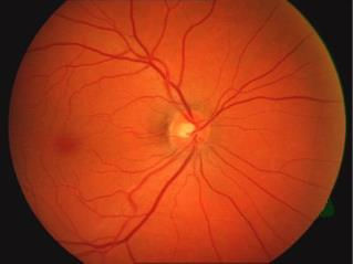 Image 1: Slit Lamp OD (Episcleral Nodule)
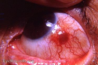 Image 2: Posterior Pole OD (Normal)
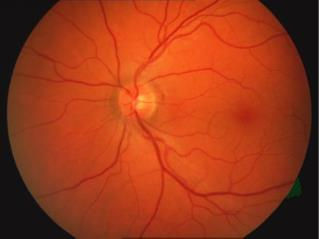 Image 3: Posterior Pole OS (Normal)
✓ Correct Option: A. Nodular episcleritis
Rationale: The localized, tender nodule is freely mobile over the underlying sclera and blanches with phenylephrine, confirming episcleritis over scleritis (which is non-mobile and does not blanch). Strongly linked to inflammatory bowel disease (Crohn's).
✓ Correct Option: A. Joint inflammation (Peripheral Arthritis / Spondylitis)
Rationale: Enteropathic arthropathy (peripheral arthritis / sacroiliitis) occurs in up to 25–30% of Crohn's patients and is the #1 most common extra-intestinal manifestation.
✓ Correct Option: E. Fluorometholone 0.1% (FML) suspension QID
Rationale: The patient's baseline IOP is already elevated at 24/25 mmHg and he is taking oral prednisone. High-potency ketone steroids (Pred Forte) carry high risk of severe secondary steroid-induced glaucoma spike. FML (soft surface ketone) has poor intraocular penetration, effectively treating surface episcleral inflammation with minimal risk of IOP spike.
✓ Correct Option: B. 4 days
Rationale: Nodular episcleritis treated with topical steroids requires re-evaluation in 3 to 7 days (4 days is optimal) to confirm resolution of inflammation, recheck Goldmann IOP for steroid spikes, and initiate weekly step-down tapering.
07. Interactive Clinical Tools
TOOLS08. Practice Case Simulator & Self-Test
EXAM MODEA 67-year-old male with newly diagnosed POAG has moderate COPD and sinus bradycardia (heart rate 50 bpm). Which is the MOST appropriate first-line pressure-lowering agent?
Non-selective beta-blockers like Timolol are absolutely contraindicated in patients with asthma/COPD and bradycardia. Latanoprost is first-line with zero pulmonary/cardiac contraindications.